We add 3D guidance to your structural heart disease therapy efficiently. syngo DynaCT Cardiac, syngo Aortic Valve Guidance, syngo TrueFusion as well as the ACUSON SC2000™ Ultrasound System with True Volume TEE provide support through a high level of automation.

syngo TrueFusion
Easy access to fusion imaging
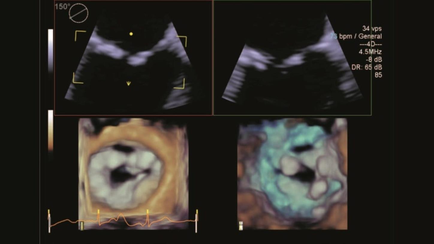
When treating structural heart disease, advanced real-time 3D TEE and True Volume color Doppler imaging are required to visualize form, function, and flow in one volume view – without compromise. With TrueFusion2, add the relevant anatomical and functional landmarks easily to your live fluoroscopy.

syngo Aortic Valve Guidance
Automated workflow and enhanced accuracy for aortic valve procedures
syngo Aortic Valve Guidance supports the interventional cardiologists and surgeons perform valve implantations. It provides fast and precise 3D information on the aortic root anatomy – making it an excellent basis for valve implantation planning.

syngo DynaCT Cardiac
CT-like imaging in the cath lab
Enhance your diagnostic and therapeutic capabilities by visualizing cardiac structures like the aortic root, the left atrium and LAA or pulmonary veins in 3D, in the cath lab. Based on rotational angiography and advanced 3D reconstruction, syngo DynaCT provides CT-like images at the time of your intervention. For optimal and reproducible results the 3D wizard intuitively guides you through the acquisition.
Clinical Content
LAA closure with fusion of live angiography and TEE imaging
At the Center for Cardiovascular Interventions Bonn, Germany, Professor Georg Nickenig and his team are performing more than 500 structural heart interventions per year. Find out what benefits they see in fusion of live angiography imaging and TEE in an example case of a left atrial appendage (LAA) closure.
High-end multimodality imaging for improved TAVI outcomes
Watch Professor Stephan Achenbach, MD, and Martin Arnold, MD, in a procedure to treat an 87-year-old patient with severe aortic valve stenosis by implanting a transcatheter aortic valve prosthesis at University Hospital Erlangen and see how they integrate imaging.
Integrated imaging solutions for structural interventions
Professor Stephan Achenbach, MD, is convinced that high-end imaging optimizes clinical results and translates into patient benefits. In unexpected situations during structural interventions in particular, continuous intraprocedural 4D TEE proved to be very helpful.
Implanting a Lotus valve in a severely stenotic mitral valve
A 75-year-old woman presented with the primary complaint of severe dyspnea at rest to Erasmus Medical Center in Rotterdam. Find out how Nicolas van Mieghem, MD, diagnosed an extensive calcification of the mitral annulus and leaflets and see the steps he took for treatment and follow-up.

Crucial imaging support with 3D TEE in LAAC
Patients with atrial fibrillation who cannot take long‐term anticoagulants have conventionally had few options for lowering their risk of stroke. Left atrial appendage closure (LAAC) – supported by advanced imaging technologies and guidance software – is an attractive alternative for these patients.

A true understanding of the patient’s heart
Stéphane Lafitte, MD, ranks among the world’s most renowned researchers in the field of echocardiography. When treating valvular diseases, real-time 3D volume color Doppler imaging provides Lafitte with information that can improve patient outcomes in cardiology.

Imaging is a vital part of a successful TAVI procedure
The global market for TAVI is expected to grow substantially and imaging will continue to play a key role in the success of TAVI procedures. Multimodality and integrative imaging approaches in particular are required to support increasingly complex, minimally-invasive structural heart interventions.