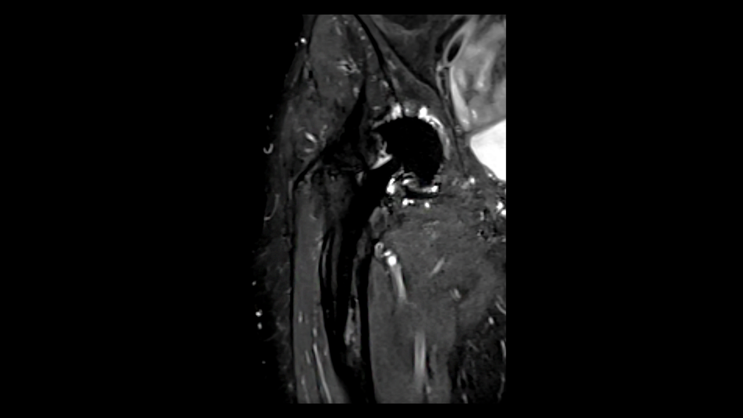
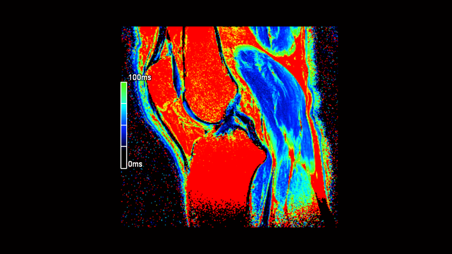
Musculoskeletal Imaging Diagnostic power of MR Imaging

Musculoskeletal Imaging

MRI is often the preferred imaging modality in musculoskeletal conditions because of its superior soft-tissue contrast, allowing the visualization of tears and degeneration. Therefore, Siemens Healthineers provides you several MRI technologies that help you achieve reliable results at exceptional speed even in the presence of challenges such as imaging of metal implants1. Achieve consistent and reproducible high-quality image results with dedicated MSK scan protocols. Gain further clinical insights from leading experts on their clinical challenges and improve your clinical routine with our latest talks.

Applications

GOKnee3D

GOKnee3D delivers reliable quality at exceptional speed. It enables a clinically validated, push-button, high-resolution 3D isotropic knee exam, with all relevant contrasts and image reading in all planes in only 10 minutes2. GOKnee3D is enabled by CAIPIRINHA SPACE and the Large Joint Dot Engine, providing push-button functionality and high consistency in knee imaging.

Your benefits at a glance:

  • Clinically validated, high-resolution, isotropic 3D knee exam in 10 minutes2
  • All essential clinical contrasts with image reading in all planes
  • Improve patient throughput and reduce costs per scan
      
Simultaneous Multi-Slice TSE

The turbo spin echo (TSE) sequence is the clinical mainstay of MSK imaging. High-resolution 2D scans typically add up to 15-20 minutes total exam time. Simultaneous Multi-Slice TSE helps cut scan times by up to 46%3 and increases slice coverage as well as resolution in routine MSK examination.
Simultaneous Multi-Slice excites several 2D slices at the same time and separates the information during reconstruction with an intelligent framework. Simultaneous Multi-Slice TSE can replace the conventional pulse sequence for almost all MSK regions.

Your benefits at a glance:

  • Reduce scan times by up to 46%3
  • Achieve higher spatial resolution and thinner slices
  • Improve patient throughput and schedule shorter exam slots per patient
     
Compressed Sensing SEMAC 2

SEMAC is especially helpful in the imaging of large metal implants1 (hip and knee arthroplasties) and helps enable critical diagnoses such as implant failure, loosening, and infection.With Compressed Sensing SEMAC, in particular, high quality imaging in the presence of MR-conditional metal implants can be performed with up to 55%3 shorter scan times. With Compressed Sensing, significant shorter acquistion times make SEMAC clinically viable.

Your benefits at a glance:

  • Imaging of MR-conditional metal implants become clinical routine
  • Robust image quality and fewer repeated scans, ensuring high patient throughput
  • Potential to diagnose clinical conditions such as prosthesis loosening, infections, inflammations, and implant failure
  • Significant shorter acquisition time without the loss of image quality
     
Large Joint Dot Engine

MR imaging of extremities is performed at 98% of MRI sites3 and constitutes 21%3 of total MRI procedures. Making these examinations easier, more reliable, and more consistent can help improve image quality while improving patient throughput for this core examination. The Large Joint Dot Engine simplifies the examination of all large joints (knee, shoulder, hip) and minimizes potential sources of planning errors with AutoAlign and AutoCoverage, powered by Artificial Intelligence. Expand your diagnostic confidence with ready-to-use optimized protocols for artifact reduction or motion robust imaging with single-click changes to a scan strategy.

Your benefits at a glance:

  • Consistent and efficient exam planning powered by Artificial Intelligence
  • At any time reproducible and high quality image results
  • Ready-to-use optimized protocols for artifact reduction or motion robust imaging
     
MapIt

MapIt provides the protocols and inline calculation of parametric maps of T1, T2, and T2* properties of the imaged tissue.

Your Benefits at a glance:

  • Moving MSK MR imaging ahead by evaluation of macromolecular structures
  • It allows the early detection of osteoarthritic pathology
  • It enables follow-up of treatment in patients with Osteoarthritis and after sports injury
     

Clinical Use

Dr. Jan Fritz

Sign up for our live webinar with Dr. Jan Fritz, taking place on May 28, 2019 at 2:00 PM (EST).

The Associate Professor of Radiology from the Johns Hopkins Hospital will give you insights on new acceleration techniques in MSK imaging such as Compressed Sensing SPACE, Compressed Sensing SEMAC, and Simultaneous Multi-Slice that help improve efficiency and the patient experience.
 

View the benefits of SEMAC

1
2
3
Subscription Button Icon
Be the first to know about our events, training, and news