Neurology

Artificial intelligence supports clinicians in reading brain images

A new software helps radiologists view, analyze, and evaluate brain images.

4min
Panagis Galiatsatos
Published on 22. Juni 2021

In Greece, as in other countries, there is an increase of neurodegenerative diseases, predominantly Alzheimer’s Disease – mainly because of the aging population.[1] Although it is the neurologist who is responsible for the diagnosis, radiology is playing an increasingly important role. This is manifest in the work of radiologist Andreas Papadopoulos, MD, PhD, scientific coordinator at Iatropolis Medical Group, who has come to appreciate the benefits of artificial intelligence.
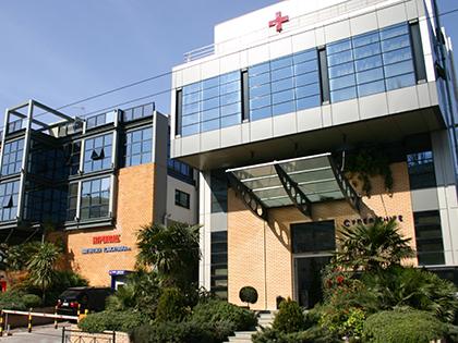
The headquarters of Iatropolis Medical Group, Greece’s leading diagnostics provider, is located in Halandri, a suburb north of Athens. The neighborhood is uncharacteristically calm because of current COVID-19 restrictions. But outside the Iatropolis building, there is a gathering of people waiting for their appointments. Most are elderly; they wait alone or accompanied by relatives.

As in many industrialized countries, the population in Greece is getting older. As a result, the Greek Ministry of Health expects a significant increase in neurodegenerative diseases. Among these, Alzheimer’s Disease is the most prevalent, accounting for 70 percent of neurodegenerative disease cases. According to estimates published by the Alzheimer Society of Greece, already 197,000 people suffer from the disease today. This number is expected to rise to 354,000 by 2050.[2]

“The likelihood of developing Alzheimer’s may be only one to two percent at age 65. But then it doubles every five years. Existing drugs cannot reverse the course of the degeneration, they can only slow it down. This is why it’s crucial to make the right diagnosis in the preliminary stages – when the first mild cognitive disorder appears -–and to filter out Alzheimer’s patients,” explains Dr. Papadopoulos.

Innovation and cutting-edge technology in medical diagnostics are a hallmark of Iatropolis, which started operating the first magnetic resonance imaging (MRI) scanner in Greece back in 1986.

Since then, Iatropolis has grown consistently and now operates seven health centers in the greater Athens area.

Iatropolis Halandri, Athens, Greece

However, the company has remained true to its ethos and continues to equip its centers with the latest technology. As of late October 2020, their inventory includes the AI-Rad Companion Brain MR1. This artificial intelligence (AI)-based software could play an important role in early diagnostics of neurodegenerative diseases.

When it comes to technical innovation, Papadopoulos is a pioneer. In 1988, when he began his radiology residency training at Aretaieion Hospital, the first MRI in the Greek public health system was installed there. In the following years, he witnessed firsthand the technological revolution that totally transformed his area of specialty. Papadopoulos has taken advantage of almost every innovation in his field and tested it in practice; he has made more than 200,000 diagnoses based on MRI and computed tomography scans. “In all these years, I gained a lot of experience and I overcame my fear of computers and all new tools,” he says.

At Iatropolis, 12,000 MRI scans of the brain are performed annually.

MRI scan of the brain

Iatropolis works with the public health system in Greece. Many of its diagnostic services are accepted and paid for by public health insurers. But not MRI scans that target neurodegenerative diseases – patients must pay for those out of their own pockets or have private insurance.

For three months now, Papadopoulos has been using AI-Rad Companion Brain MR for these examinations. “This is a brain volumetry software,” he explains. “That means it provides automatic volumetric quantification of different brain segments. It is able to separate them from each other: It isolates the hippocampi and the lobes of the brain and quantifies white matter and gray matter volumes for each segment separately.”

But would he not be able to make these calculations himself? “Absolutely not,” says Papadopoulos. It involves a tremendous amount of work and, above all, a level of precision that humans simply cannot achieve, he says. But more than that, thanks to the software, he can compare the results with normative data from a healthy population:


Andreas Papadopoulos

“This is especially helpful when the radiologist questions if what he recognizes as volumetric change is really volumetric change and not perhaps a normal sign of aging.”

These possibilities opened up by AI are what Papadopoulos has in mind when he talks about early detection of neurodegenerative diseases. “In the early stages, the volumetric changes are small. In the hippocampus, for example, there is a volume reduction of 10 to 15 percent, which is very difficult for the eye to detect. But the objective calculations provided by the system could prove a big help.”

As a radiologist, Dr. Papadopoulos does not normally come into contact with patients. He performs his examination in the laboratory and on the screen. However, he would be available to any patient who would want to see him. I ask him whether he tells the patients that they have neurodegenerative disease? Papadopoulos shakes his head: “I do not report to a patient that he or she has, for example, Alzheimer’s, because this is not a radiological diagnosis. That is the neurologist’s job. I also do not mention the word Alzheimer’s in my report. I describe the volumetric changes and the neurologist interprets the findings in association with the results of neuropsychological testing.”

AI aims to relieve physicians from a lot of work and, if properly embedded in the workflow, to save time. However, this is not the case for Dr. Papadopoulos – at least not yet. During the initial phase, he says the software is more likely to mean more work for him: “I’m currently singling out two to three cases a day and evaluating them with AI-Rad Companion Brain MR.” Mostly, these are completely normal cases or are cases in the borderline range where you cannot be absolutely sure if there is a volumetric change. “In the first group I want to validate the results of AI. In many cases of the second group, I use the results of the software and incorporate them into my diagnosis.” Later, once his new workflow is established, he will find out if AI also saves him time.

Already, however, Papadopoulos recognizes that his biggest benefit is “the software providing an objective framework on which I can base my subjective assessment during an examination.”

A big benefit is the software providing an objective framework on which one can base a subjective assessment during an examination.

Another promising field of application for AI is the research into drugs and new treatments for neurodegenerative diseases. Says Dr. Papadopoulos: “again, the changes in brain volumes during treatment are gradual and mild; the human eye cannot perceive them. AI, however, can show whether the patient’s condition continues to deteriorate or whether new drugs are able to prevent further degeneration. This introduces objective parameters for evaluating the therapy.”


By Panagis Galiatsatos
Panagis Galiatsatos is a newspaper and radio journalist based in Athens, Greece, and is familiar with the field of health economics, among others.