


Oncology
Searching for the causes of cancer
In addition to investigating the causes, researchers are motivated by one thing above all else: the search for a cure.
4min
Published on July 30, 2021
Over 19 million cases worldwide each year – and the number is rising. Some people are affected in old age, others in childhood. Whenever cancer strikes, it is a life-changing disease. Its exact origins have been the subject of research since the advent of modern medicine over 150 years ago. As well as investigating the causes, researchers are motivated by one thing above all else: the search for a cure.
The birth of radiotherapy


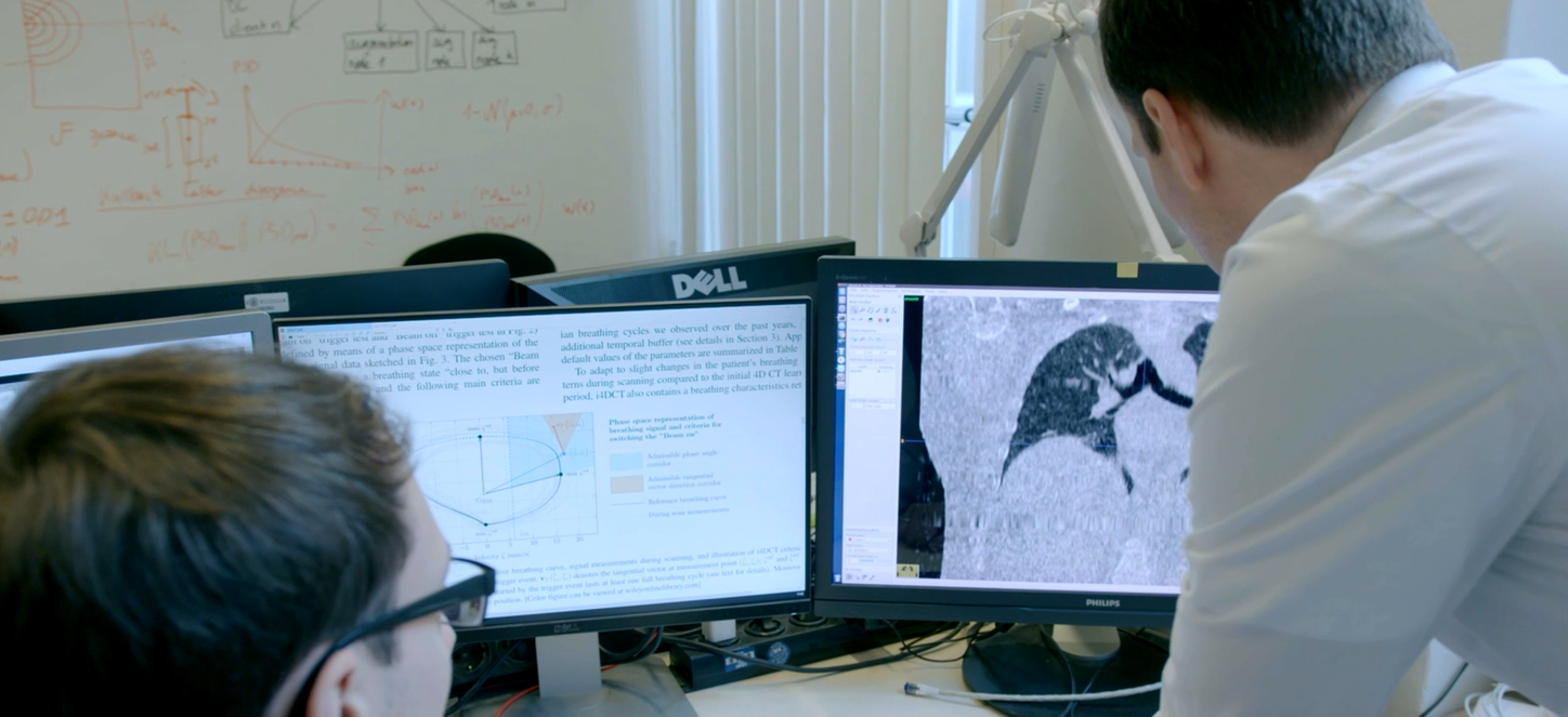
Computed Tomography imaging for radiation therapy planning of an otolaryngologic tumor together with MR and PET-imaging. (Courtesy of University Hospital Erlangen, Germany)


- References