Imaging

Anatomical lectures with Cinematic Anatomy 

An Austrian university takes the teaching of anatomy to the next level. At the same time this saves costs and resources in the long term.

6min
Lena Stauber
Published on September 13, 2022

Have you ever consulted a medical textbook and tried to grasp its anatomical illustrations? Medical teaching is extremely complex—but technology can help. Cinematic Anatomy1 is an immersive application that helps medical students, medical professionals, and patients better understand the human body.

Students at an Austrian university are learning about anatomy in stereoscopic 3D—thanks to a venue developed by the Ars Electronica Futurelab. The medSPACE auditorium at Johannes Kepler University (JKU) in Linz allows lecturers such as Professor Franz Fellner, MD, Head of Radiology at Kepler University Hospital, to display human anatomy in images that are almost indistinguishable from photographs. These virtual flights into the human body are made possible by Cinematic Anatomy, an application based on Cinematic Rendering.
Cinematic Anatom, an application based on Cinematic Rendering
3D renderings are already being used in imaging. The standard method is known as volume rendering and is common in the film industry. Cinematic Rendering involves passing light from a virtual source through an image dataset to simulate the transparency and color properties of the tissue and create the illusion of a 3D object. It uses a complex illumination model that incorporates many of the light-scattering, absorption, and shading effects that are seen when viewing things in everyday surroundings [1]. This makes the images extremely realistic and simplifies things such as surgery planning and anatomical understanding.
Cinematic Anatomy
Cinematic Anatomy was developed in collaboration between the JKU and Siemens Healthineers. The aim was to take the teaching of anatomy at medical universities and hospitals to the next level. It is currently being used in Linz in a world-first. When the Faculty of Medicine was planning new premises, the idea of teaching anatomy virtually was mooted as a way of saving costs and resources in the long term. The infrastructure required to prepare corpses for dissection is expensive and takes up a great deal of space.

Cinematic Anatomy has a feature called Stereoview, which allows audiences to view the photorealistic renderings in 3D using 3D glasses. 
Fellner, who was involved in the collaboration and has invested a number of years in research, is enthusiastic about the opportunities: “Cinematic Anatomy is a fantastic technology that produces incredible, razor-sharp images of the human anatomy. It provides a cinematic tour of the body. This is real cross-sectional anatomy studied on real bodies, not in simulated reality as is the case with computer graphics programs.”

The benefits also extend beyond medical teaching: “Patients themselves benefit from Cinematic Anatomy, as 3D visualizations are much easier to understand than standard CT or MRI images. And it’s naturally always to their advantage when the attending physicians have enjoyed the best possible training,” says Andrea Hofbauer, Collaboration Manager at Siemens Healthineers. She played a key role in developing the Cinematic Anatomy prototype in Princeton, USA.

Andrea Hofbauer, Collaboration Manager at Siemens Healthineers, Princeton, USA
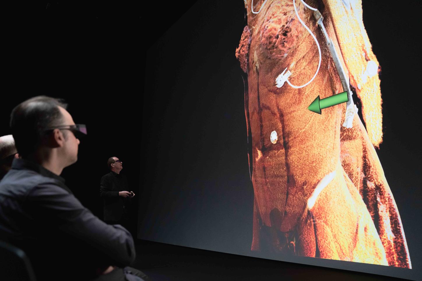
Cinematic Anatomy can also virtualize the various structures of the body through special windowing tools separating high contrast like bones and contrast filled vessels, from soft tissue. With Clip planes, carve and crop tool structures of interest can get segmented like in virtual dissection.
However, Cinematic Anatomy does not replace practical work on the human body in the dissection room. Medical students in Linz still need to learn to dissect real bodies, and they do so in a four-week course held in Graz. This is topped up by multiple weekly sessions of the new anatomy class with its Cinematic Anatomy images. The 3D images are invaluable when it comes to providing a clear view of the structure of the human body so that dissection can be planned in detail.
The new application has also made anatomy lectures more sustainable and modern. “
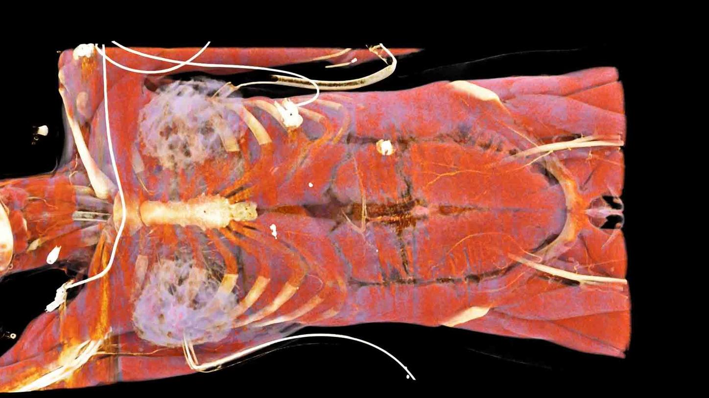
Not only can we enlarge our images, but we can also move them, rotate them, and virtually cut into the complex 3D anatomy, making it much easier to understand. Moreover, it’s a nondestructive process. What we cut off a corpse is gone forever, but we don’t have to be afraid of this in virtual anatomy,” explains Fellner, and adds that “the viewer has a totally different relationship with the images knowing that this is a living person.”

Nominated for the 2017 German Future Prize: Professor Franz Fellner, MD

Looking ahead, there are plans to combine Cinematic Anatomy with augmented reality— and to develop a Cinematic Anatomy community whose members will build up a content library to create a complete digital atlas of the human anatomy. As Professor Fellner believes: “This is just the beginning. It has incredible potential.”


By Lena Stauber

Lena Stauber is an editor in corporate communications at Siemens Healthineers. The team specializes in topics related to healthcare, medical technology, disease areas, and digitalization.